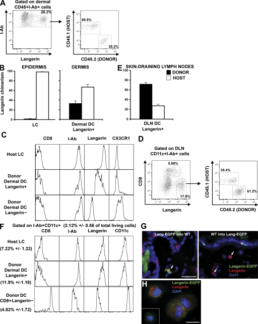
Figure 4.
Homeostasis of langerin+ DCs after congenic BM transplantation. Lethally irradiated host CD45.2+C57BL/6 were transplanted with congenic donor CD45.1+C57BL/6 BM (CD45.1 into CD45.2 chimeric mice). 2 mo after reconstitution, host versus donor chimerism was measured in the langerin+ DC population in the skin (A and B) and in skin DLNs (D and E). (A) Dot plots show I-Ab and langerin expression among dermal CD45+ cells, and the host versus donor chimerism of langerin+ dermal DCs (CD45+I-Ab+). (B) Mean values of langerin+ DCs chimerism in the dermis. LC (CD45+I-Ab+ langerin+) chimerism in the epidermis is shown as control. (C) Expression of different markers was compared by flow cytometry between host remaining (CD45.2+I-Ab+ langerin+) LCs and donor-derived langerin− or langerin+ dermal DCs. (D) Dot plots show CD8 versus langerin expression among CD11c+I-Ab+ cells in the skin DLNs and host versus donor chimerism of CD8− langerin+ DCs. (E) Mean values of langerin+ DC chimerism. (F) Phenotype of donor-derived CD8− DC langerin+, CD8+ DC langerin−, and host remaining LCs. (G) Back skin cross-sections isolated from CD45.2 Langerin-EGFP into CD45.1 chimeric mice (left) or the reverse CD45.1 into CD45.2 Langerin-EGFP chimeric mice (right) were stained with anti-langerin antibody. Nuclei were counterstained with DAPI. Bar, 50 μm. (H) Dermal cell suspensions were prepared from CD45.2 Langerin-EGFP into CD45.1 chimeric mice. Donor langerin+ dermal DCs were sorted on the criteria of CD45.2, I-Ab, and EGFP expression and harvested onto cytospin slides for langerin staining. Inset shows langerin staining control. Bar, 10 μm. Representative data from four independent experiments are shown. Each experiment included at least three separately analyzed mice; error bars represent the SD between the results obtained from each of the three mice.

