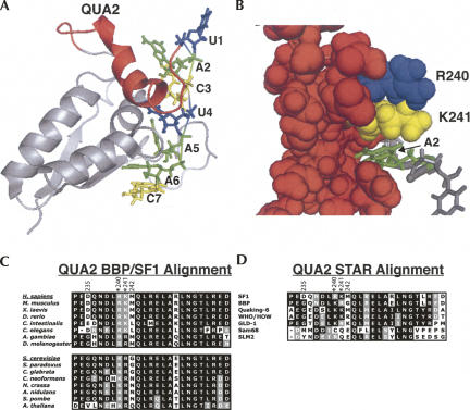
FIGURE 8.
Analyzing the QUA2 domain structure from the NMR structure of SF1 in complex with the branchpoint sequence RNA (PDB #1K1G) (Liu et al. 2001) and alignments of branchpoint binding proteins from multiple organisms and the related STAR/GSG proteins. (A) The QUA2 domain (red) relative to the KH domain and branchpoint sequence. The nucleotides of the branchpoint sequence are numbered 1 through 7. (B) A spaced-filled view of R240 (blue) and K241 (yellow) and the rest of the QUA2 domain (red) binding RNA. The A2 nucleotide (green) fits in a pocket formed by the QUA2 domain. (C) An alignment of QUA2 domains of SF1, BBP, and branchpoint binding proteins from different organisms. (D) A QUA2 domain alignment of SF1, BBP, and other STAR/GSG proteins. Accession numbers for these proteins can be found in Supplemental Table 1.

