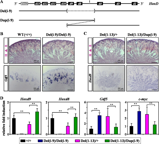
Figure 8. Deletion of Both Hoxd8 and Hoxd9 Leads to Epithelial Microcyst Formation.
(A) Scheme showing the intact HoxD cluster (top) and two newly produced mouse strains used to characterize Hoxd genes responsible for microcyst formation. The strains are referred to as Del(i-9) or Dup(i-9) for a deletion or a duplication, respectively, and they span from ‘region i' to Hoxd9, inclusively (dashed lined).
(B, C) Kidney cryosections of WT (+/+), Del(i-9) homozygous (Del(i-9)/Del(i-9), Del(1–13) heterozygous (Del(1–13)/+) and Del(1–13)/Dup(i-9) compound mice were either stained with H&E (top), or processed for the detection of Gdf5 (left) or Hoxd9 (right) by ISH. The Del(1–13)/Dup(i-9) compound strain was obtained by breeding Del(1–13) and Dup(i-9) heterozygous mice.
(D) qPCR was used to compare the expression levels of Hoxd9, Hoxd8, Gdf5 and c-myc in newborn kidneys from the mouse strains described before. Values represent the mean of three independent experiments, after normalization to the WT. Independent Student's t test: **, P < 0.01.

