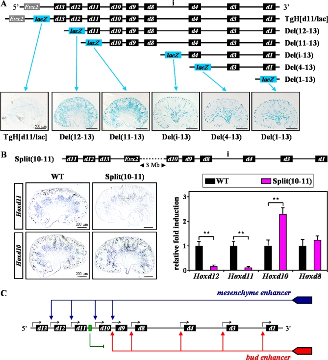
Figure 9. Searching for Putative Mesenchyme and Ureteric Bud Enhancers.
(A) Schematic representation of a panel of in vivo HoxD alleles containing the Hoxd11/lacZ fusion reporter gene together with progressive deletions of the cluster. Blue boxes (lacZ) indicate the position of the same reporter transgene at the posterior end (5', left) of the intact (TgH[d11/lac]) or deleted (Del) HoxD cluster. Representative stainings of newborn kidneys are shown below.
(B) Scheme of the inversion-mediated split of the HoxD cluster between Hoxd11 and Hoxd10 (top; Split(10–11)) [23], separating the two half-clusters by approximately 3 Mb. ISH for Hoxd11 and Hoxd10 genes (left), as well as qPCR for Hoxd12, Hoxd11, Hoxd10 and Hoxd8 (right) were carried out in WT and Split(10–11) specimen. Independent Student's t test: **, P < 0.01.
(C) Model of global Hoxd gene regulation during kidney development. The different expression domains are likely controlled by regulatory elements localized outside the HoxD complex, at the telomeric (3') side. The bud enhancer may control the expression of anterior genes (from Hoxd9 to Hoxd1) in the UB (red arrows). Posterior Hoxd genes are not responsive to this enhancer, because of the presence of a putative silencer or boundary element (green arrow), located between Hoxd10 and Hoxd11. The mesenchyme enhancer may trigger the expression of posterior genes (from Hoxd12 to Hoxd9) in the MM.

