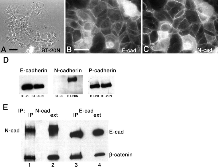
Figure 4.
Expression of N-cadherin by BT-20 cells. BT-20 cells were transfected with N-cadherin (BT-20N) and expression induced with dexamethasone. A, Phase-microscopy of living BT-20N cells. Bar, 10 μm. B and C, Cells were grown on glass coverslips and processed for coimmunofluorescence localization with antibodies against E-cadherin (Jelly; B) and N-cadherin (C). D, BT-20 and BT-20N cells were extracted with NP-40 and 20 μg protein from each extract was resolved by SDS-PAGE, transferred to nitrocellulose, and immunoblotted for E-cadherin (HECD1), N-cadherin, or P-cadherin. E, Extracts of BT-20N cells were immunoprecipitated with antibodies against N-cadherin or E-cadherin (HECD1). The immunoprecipitation reactions, as well as cell extracts, were resolved by SDS-PAGE, transferred to nitrocellulose, and immunoblotted for N-cadherin and β-catenin (lanes 1 and 2) or E-cadherin (HECD1) and β-catenin (lanes 3 and 4).

