
Figure 3.
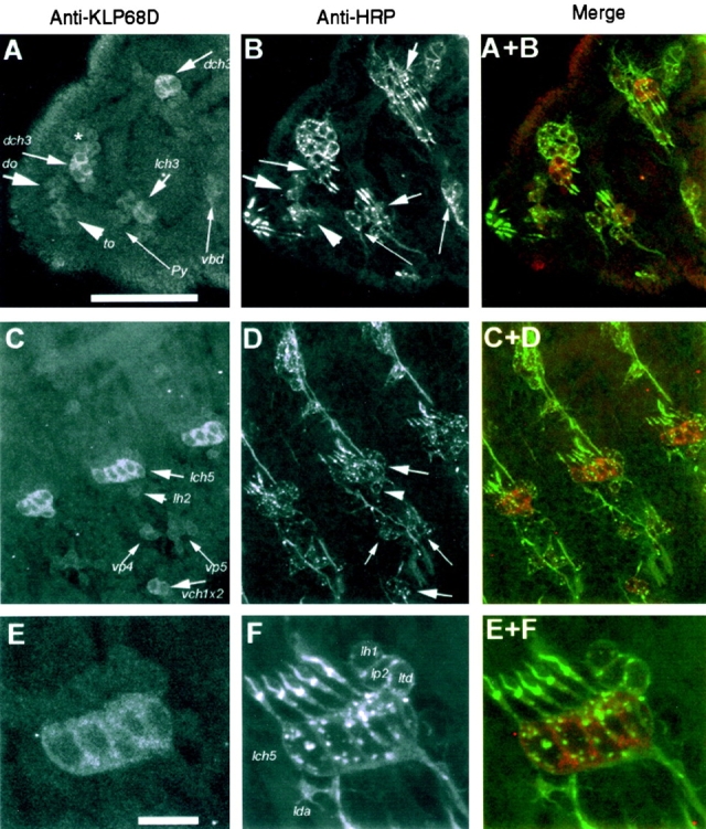
Immunostaining pattern of rabbit anti-KLP68D (A, C, and E) and anti-HRP (B, D, and F) in the epidermis of a late stage 16 embryo. The anterior side of the embryo in all the figures is placed towards the left and the dorsal side is upwards. The KLP68D antiserum stained neuronal soma of a subset of sensilla while anti-HRP marked all the sensory neurons in the epidermis (Jan and Jan 1982). Sense organ nomenclature is followed as in Campos-Ortega and Hartenstein 1997. Bars: A, 50 μm (A–D); E, 10 μm (E and F). (A) The anterior part of the embryo is shown, including the first two thoracic segments. Relatively strong staining of the dorsal (dch3) and lateral (lch3) chordotonal organ neurons in the first and second thoracic segments is evident. The polyinnervated external sense organ (vbd) and external papilla (Py) in the second and first thoracic segments, respectively, are only weakly stained. Similarly, the dorsal (do) or antennalles organ and the terminal organ (to) or maxillarorgan are weakly stained. Most of the sensory cells of these two organs are just beyond the plane of focus. The faint staining observed in a group of cells (indicated by an asterisk) in the dorsal region of the first thoracic segment are identified as bolwig organ. (B) The same region as in A labeled with goat anti-HRP, which stains all neuronal cell bodies and processes and clearly shows that only a subset of neurons are stained for KLP68D. (C) Staining in the abdominal segments is shown. The cell bodies of the lateral (lch5) and the ventral (vch1x2) chordotonal neurons are relatively strongly stained. The neuronal cell bodies of ventral papillae (vp4 and vp5) and the lateral trichoid sensillum (lh2) are less intensely stained. In addition, there is weak staining in some unrecognized cells in between vp4 and vp5. (D) Same field as in C stained with anti-HRP. (E) A high magnification image of a region of the lateral epidermis containing the pentascholopedial organ (lch5) and other sensilla. Staining is restricted to the neuronal cytoplasm of the lch5. (F) The same field as in E stained with anti-HRP, which highlights all the neurons. Identified neurons are marked in the figure. The neurons of the monoinnervated external sensilla, lh1 and lp2, and the multi-dendritic neurons, ltd and lda, are not stained significantly relative to lch5.