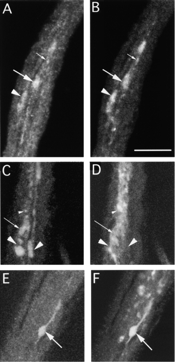
Figure 4.

Segmental nerve bundles from a Klc1/Df(3L)8ex94 third instar larva, simultaneously stained with either mouse anti-ChAT (A) and rabbit anti-KLP68D (B) or with anti-ChAT (C and E) and rabbit anti-SYT (D and F). They are visualized using FITC anti–mouse and Cy5 anti–rabbit. Both frames are simultaneously excited and captured by separate photomultipliers in a single optical scan of 1-μm thickness. Accumulations of the respective antigens in the axons in individual clogs are marked by different sized arrows and arrowheads. Both KLP68D and ChAT antisera usually stain the same foci (clogs) in the nerve roots (see Table for details). SYT generally does not associate (arrowheads in C and D) with the ChAT in these foci although occasional coincident staining is seen (arrows in E and F). Bar, 10 μm.
