
Figure 6.
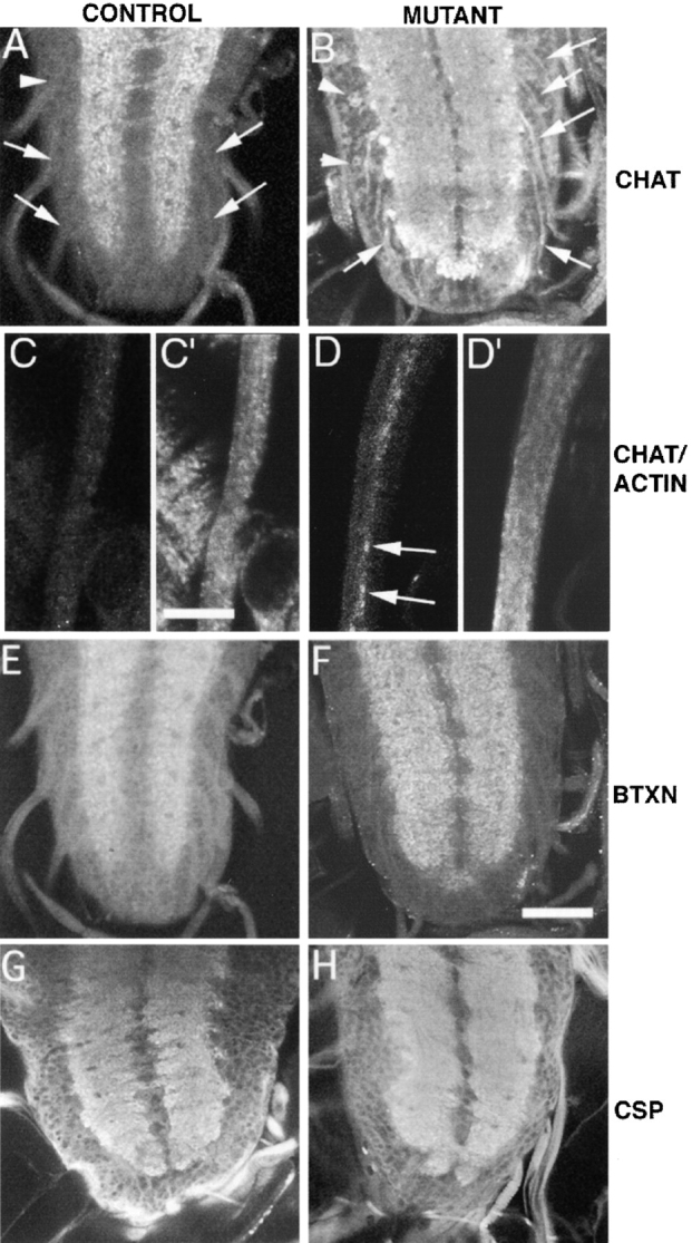
Staining of various markers of axonal transport in the cell bodies and axons of heterozygous (A and C) and homozygous (B and D) klp64D mutant larval brains and segmental nerves. Samples are stained with mouse anti-ChAT (A–D), XTRITC-coupled α-bungarotoxin (E and F) and with mouse anti-CSP antibody (G and H), respectively. A few were also stained with rabbit antiactin antibody (C′ and D′). Bars: C′, 10 μm (C, C′, D, and D′); F, 100 μm (A, B, E–H). (A) Klp64Dk1/TM3 third instar larva ventral ganglion has anti-ChAT staining confined to the neuropil. The punctate nature of the staining indicates localization of ChAT in synaptic bulbs. There was no staining in the cell cortex (arrowhead) or in the nerve roots (arrows). (B) The homozygous Klp64Dk1third instar larval brain exhibited significant ChAT staining in the axons (arrows) of the nerve roots and in the cell cortex (arrowheads). Abnormally strong staining can also be seen at the boundary of the neuropil. (C and C′) A part of a segmental nerve bundle is shown from a Klp64Dk1/TM3 larva stained with mouse anti-ChAT and rabbit antiactin antibody, respectively. Both cases showed no specific accumulation of these two antigens in any of the axons. (D and D′) The equivalent part of a similar nerve bundle from a Klp64Dk1 homozygous larva showed strong accumulations of ChAT antigen in some of the axons (arrows). The antiactin staining in the axons is slightly higher in intensity than the control, but it is not selectively higher in the axons that showed accumulated ChAT as shown in F. E–H show equivalent regions of ventral ganglia from Klp64Dk1/TM3 (E and G), and the Klp64Dk1 homozygous third instar larvae, respectively. They are stained with α-bungarotoxin (E and F) and anti-CSP (G and H) and the pattern is similar between the control and mutants.