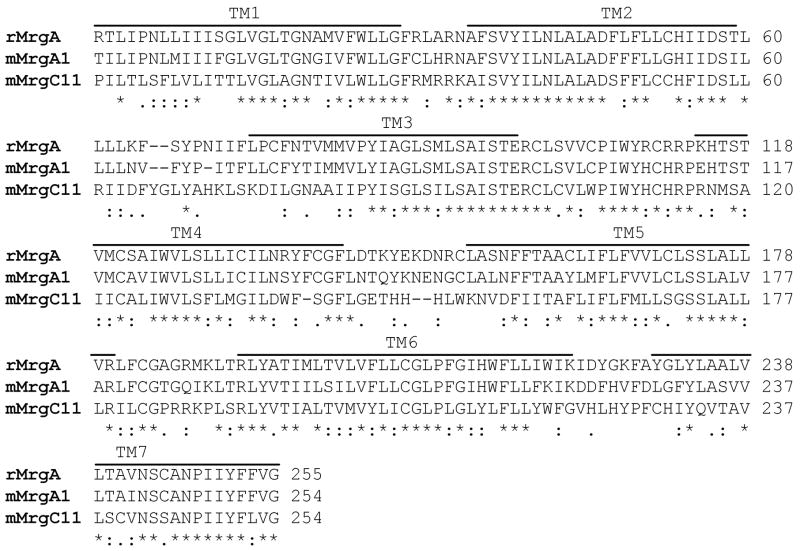
Figure 1.
The sequence alignment provided as an input for the homology modeling of rMrgA. The transmembrane regions were obtained from the mMrgC11 and mMrgA1 structures. The N-terminus (11 residues) and C-terminus (38 residues) were omitted because for class A GPCRs (rhodopsin-like) they generally do not play a role in the binding of small ligands [45].

