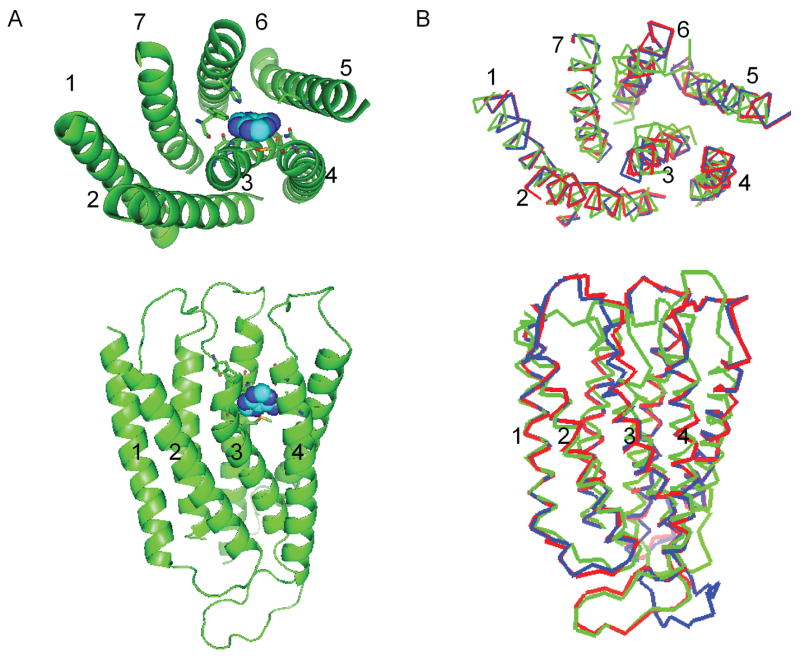
Figure 3.

The predicted 3-D structure of the rMrgA receptor. The top part shows the view from the extracellular side, while the bottom part shows the side view (with the extracellular part on top).
A) Adenine (in spheres) is docked in the rMrgA receptor. The residues within 5 Å of adenine are shown as sticks.
B) The rMrgA receptor (red) is overlapped with mMrgA1 (blue) and mMrgC11 (green).
