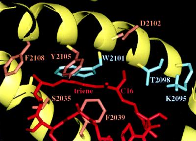
Figure 4.
X-ray structure of the FRB–rapamycin interface. Rapamycin is red and the FRB protein backbone is yellow. Some of the FRB residues selected for mutation are indicated as blue and brown; residues that led to compensatory binding to C16-(R)-isopropoxy rapamycin 3R are blue. In the first round of selection, libraries were constructed at positions F2039, T2098, W2101, D2102, Y2105, and F2108. In the context of T2098L or T2098I, libraries were constructed at S2035, F2039, K2095, W2101, and D2102 for the second round of screening. For the third generation of libraries, positions L2031, A2034, S2035, Y2038, F2039, M2047, L2051, K2095, D2102, Y2104, Y2105, and F2108 were randomized in the context of T2098L, W2101F or T2098L, W2101L.

