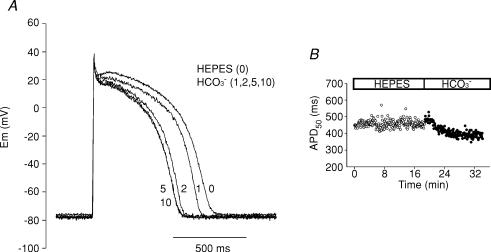
Figure 1.
APD shortening after switching the extracellular solution from Hepes to HCO3−-buffered solution A, AP recordings under current-clamp mode before (Hepes) and after 1, 2, 5 and 10 min of superfusion of a cat ventricular myocyte with external HCO3−. The presence of HCO3− in the external solution produced a gradual APD shortening. B, typical time course of beat-to-beat APD50 measured before and after switching the extracellular solution from Hepes to HCO3−.

