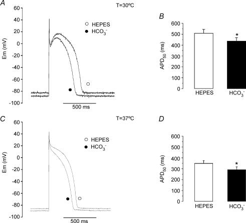
Figure 9.
APD shortening induced by external HCO3− at 30°C and 37°C A, AP recordings before (Hepes) and after 10 min of superfusion of a cat ventricular myocyte with external HCO3− at 30°C. B, average APD50 (n = 8) measured before and after 10 min of HCO3−-buffered solution at 30°C. At 30°C, HCO3− induced an APD shortening of 14.2 ± 1.6% (n = 8). C, AP recordings before (Hepes) and after 10 min of superfusion of a cat ventricular myocyte with external HCO3− at 37°C. D, average APD50 (n = 4) measured before and after 10 min of HCO3−-buffered solution at 37°C. At 37°C, HCO3− induced an APD shortening of 16.1 ± 2.3% (n = 4).

