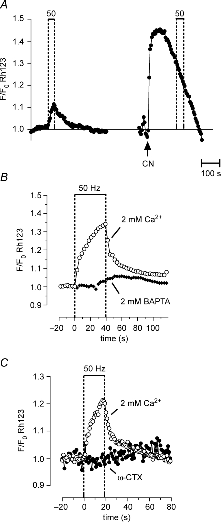
Figure 3.
Repetitive stimulation produces a partial, reversible depolarization of Ψm that depends on Ca2+ influx into motor terminals Ψm depolarization was indicated by an increase in Rh123 fluorescence (due to unquenching). A, Rh123 fluorescence changes in response to a 50 Hz, 40 s stimulus train (left), followed by addition of 1 mm cyanide (CN, right). A second 50 Hz stimulus train applied following CN exposure produced no further increase in fluorescence. The CN-induced fluorescence increase probably underestimated the CN-induced depolarization of Ψm, because of loss of Rh123 from the terminal via diffusion into the axon and bath (Nicholls & Ward, 2000). B and C, the stimulation-induced increase in Rh123 fluorescence was inhibited by adding 2 mm BAPTA (B) or 3 μmω-conotoxin GVIA (ω-CTX) to the physiological saline (C). The illustrated stimulation-induced partial Ψm depolarization was not detected in a previous study (David, 1999) that used a less-sensitive imaging system. A–C were recorded from different terminals; n = 1 experiment for A; n = 2 experiments for B and C.

