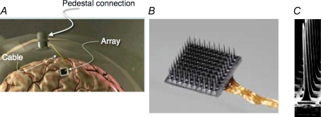
Figure 1.
NIS implant and sensor A, parts of the implant include the array, skull mounted percutaneous pedestal, and a 96 wire cable that connects them. B, 10 × 10 array of electrodes, each separated by 400 μm. C, scanning electron micrograph one electrode showing its shape and pointed, platinum (Pt)-coated tip.

