
Figure 1.
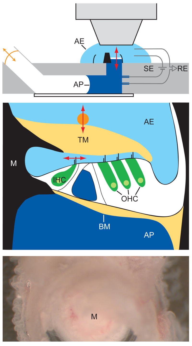
In vitro cochlear preparation. (a) The excised middle cochlear turn, shown in black as transected through the modiolus (center) and outer bony wall, separates the two compartments of the experimental chamber. The apical and basal aspects of the organ of Corti (white) are immersed in artificial endolymph (AE) and artificial perilymph (AP), respectively. Pairs of recording electrodes (RE) and stimulating electrodes (SE) measure microphonic potentials and provide transepithelial electrical stimuli. Acoustic stimuli from an earphone (orange arrow) are delivered to the basilar membrane (red arrow) through the air-and-fluid-filled lower compartment. (b) A schematic drawing of the cochlear partition as mounted in the in vitro recording chamber shows the organ of Corti (white) suspended between the bone (black) of the modiolus (M) and outer cochlear wall. The hair bundles of inner (IHC) and outer (OHC) hair cells are stimulated by shearing motions between the basilar membrane (BM) and tectorial membrane (TM). Radial movement of hair bundles of the inner hair cells (horizontal red arrow) is measured with a photodiode; vertical movement (vertical red arrow) was detected with laser interferometry using a glass bead (orange) atop the tectorial membrane. (c) A micrograph taken through a dissecting microscope displays the upper surface of the preparation. Three rows of outer hair cells spiral around the cochlear modiolus. (d) A video micrograph shows the hair bundles of inner hair cells, one of which is marked (red arrow) to indicate the axis of movements detected with a dual photodiode. (e) A similar micrograph shows two of the three rows of outer hair cells and their V-shaped hair bundles. Scale bars: (c), 100 μm; (d) and (e), 10 μm.