
Figure 2.
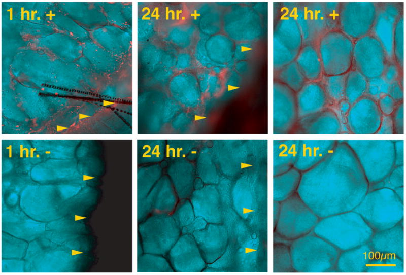
Detection of LM-332 using immunofluorescence of whole mount wounds. Wounds treated with soluble biotinylated LM-332 and saline control at 1 and 24 hours. Arrowheads indicate wound margin. In soluble biotinylated LM-332 treated wounds, bright orange staining (fluorescent emission of streptavidin-Cy5) can be seen in the wound bed at both 1 and 24 hours after application, while immunofluorescence is absent in control wounds.