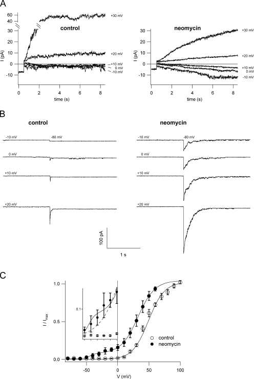
Figure 2.
Neomycin shifts the voltage dependence of SV activation. (A) Traces showing SV current activation; compared to control conditions (left) neomycin (1 mM; right) activates SV channels at more negative potentials, giving rise to inward cation currents. Currents were recorded from a large cytosolic side-out vacuolar patch in bath and pipette solutions containing 100 and 200 mM K+, respectively. (B) The threshold of SV current activation in control (left) and neomycin (1 mM, right) was probed by monitoring the deactivation tail currents upon application of the nonpermissive potential of −80 mV after a prepulse stimulus ranging from −10 to +20 mV in 10-mV steps. (C) Normalized instantaneous tail currents (see MATERIALS AND METHODS) were plotted as a function of the activating potential and fitted with a single (control; open circles) or two (1 mM neomycin; closed circles) Boltzmann functions. Data points represent the mean of seven experiments in control and three experiments in 1 mM neomycin, error bars represent SEM. Single experiment data were fitted with a Boltzmann function, normalized to the saturating current level, and averaged. In the inset, data points between −50 and 0 mV are plotted with an expanded y scale, in order to point out the “shoulder” in the normalized current data for neomycin deviating from the theoretical single Boltzmann function.

