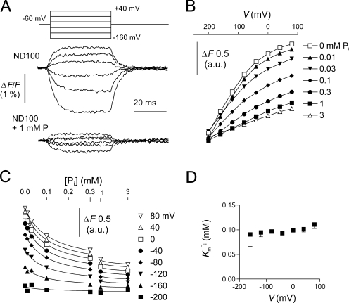
Figure 5.
Pi dependency of the voltage-dependent fluorescence in oocytes expressing S448C. (A) Original fluorescence trace recorded in ND100 (top) and ND100 + 1 mM Pi (bottom) solutions from an oocyte labeled with MTS-TMR. The membrane voltage was stepped from V
h = −60 mV to voltages in the range −200 to +80 mV in 40-mV increments, as indicated. Data were lowpass filtered at 500 Hz. (B) Pi dependency of the voltage-dependent fluorescence (ΔF). Steady-state fluorescence at different membrane potentials was acquired for each Pi concentration indicated in the figure. Data points are joined for visualization only. (C) Data in B were replotted as a function of the Pi concentration and fitted with Eq. 4 (solid lines). (D)  , as reported by the fit of Eq. 4 to the data in C (H constrained to 1).

