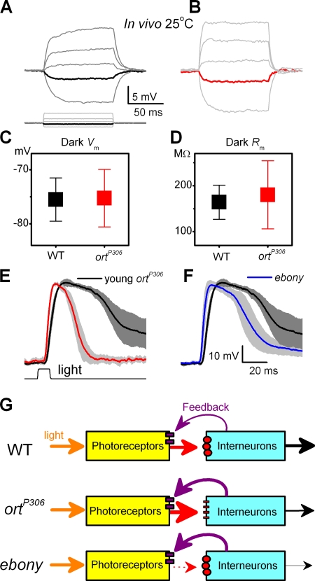
Figure 5.
Evidence that the photoreceptor output is modulated by synaptic feedback. Voltage responses of dark-adapted WT (A) and ortP306 (B) photoreceptors to small hyperpolarizing and depolarizing current pulses in current-clamp recordings in vivo. Responses to a −0.01 nA pulse is shown in black (WT) and in red (ortP306) in the respective figures. Notice that the depolarizing voltage responses of ortP306 photoreceptors are larger and peak earlier than those of WT photoreceptors, similar to our findings with light pulses in Fig. 2. (C) Resting potential of WT and ortP306 photoreceptors in darkness (mean ± SD, n = 7). (D) Impedance of the photoreceptors to −0.01 nA (mean ± SD, n = 7). The use of small negative current steps prevents the activation of voltage-gated ion channels (Hardie, 1991a). In both photoreceptors the capacitive voltage charge dies out before the end of the step, at which point the voltage is read and the impedance calculated. Responses of (E) a young ortP306 (n = 4, red, normalized), (F) adult ebony (n = 5, blue), and WT photoreceptors (n = 5, black) to a bright pulse. Mean ± SD shown. ebony mutant has a greatly reduced histamine recycling (Borycz et al., 2002), reducing the probability of successful synaptic transmission from photoreceptors to the primary visual interneurons. (G) Graphical representation of the experimental findings. Photoreceptor output is boosted when the histamine receptors (small red circles) on the interneurones malfunction (ortP306) or when there is a reduction in histamine release (dotted arrow) from the photoreceptor terminals (ebony). Both conditions can be explained by enhanced synaptic feedback from interneurones (thick purple arrows) to photoreceptors.

