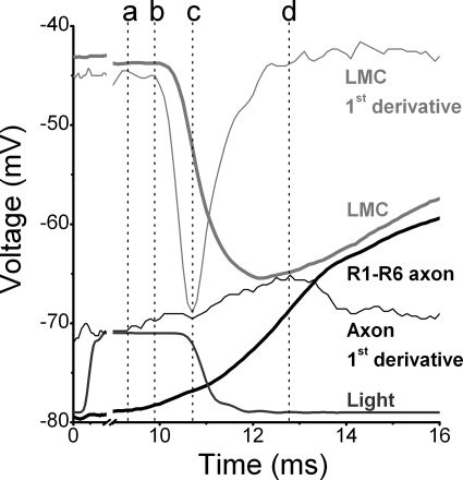
Figure 9.
Comparison of pre- and postsynaptic waveforms, recorded from lamina of WT flies at 25°C. High-quality voltage responses of an R1–R6 photoreceptor axon (black thick trace) and an LMC (light gray thick trace) and their corresponding first derivatives (thin traces of respective colors) to a 10-ms bright light pulse (18,500 photons; dark gray trace). Notice the x-axis break. The photoreceptor begins to depolarize 9 ms after the light onset (marked by dotted line at a) followed by initiation of a transient hyperpolarization in the LMC (b). When the rate of LMC hyperpolarization is at its fastest (c), the rate of photoreceptor depolarization reaches its local minimum. The photoreceptor depolarizes at its fastest rate (d) soon after LMC has begun to depolarize.

