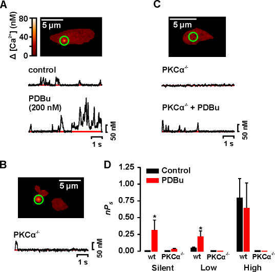
Figure 5.
PKCα is required for Ca2+ sparklets in arterial smooth muscle. Sample images of typical WT (A) and PKCα−/− (B and C) mouse arterial myocytes. The traces below the images (A and C) show the time course of [Ca2+]i in the sites marked by the green circle before and after the application of 200 nM PDBu. (D) Bar plot of the mean ± SEM nPs under control conditions and after application of PDBu. *, significantly different from control.

