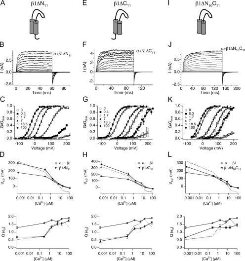
Figure 7.
Intracellular domain deletions of β1 eliminate the leftward shift of the G-V relationship at high Ca2+. (A) Cartoon of the β1ΔN11 mutant. (B) Families of BK/α+β1ΔN11 currents evoked by 60-ms depolarizations in 7 μM Ca2+. (C) Normalized G-V relationships (mean ± SEM) of BK/α+β1ΔN11 at indicated Ca2+ (n = 5–18). (D) V1/2-Ca2+ and Q-Ca2+ relationships (mean ± SEM) for BK/α+β1ΔN11 compared with BK/α and BK/α+β1. (E) Cartoon of the β1ΔC11 mutant. (F) Families of BK/α+β1ΔC11 currents evoked by 90-ms depolarizations in 7 μM Ca2+. (G) Normalized G-V relationships (mean ± SEM) of BK/α+β1ΔC11 at indicated Ca2+ (n = 4–26). (H) V1/2-Ca2+ and Q-Ca2+ relationships (mean ± SEM) for BK/α+β1ΔC11 compared with BK/α and BK/α+β1. (I) Cartoon of the β1ΔN10ΔC11 mutant. (J) Families of BK/α+β1ΔN10ΔC11 currents evoked by 150-ms depolarizations in 7 μM Ca2+. (K) Normalized G-V relationships (mean ± SEM) of BK/α+β1ΔN10ΔC11 at indicated Ca2+ (n = 3–14). (L) V1/2-Ca2+ and Q-Ca2+ relationships (mean ± SEM) for BK/α+β1ΔN10ΔC11 compared with BK/α and BK/α+β1.

