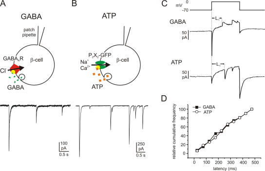
Figure 1.
Quantal GABA and ATP release in rat β cells monitored by overexpression of the ionotropic membrane receptors GABAA and P2X2. Examples of GABA- (A) and ATP-activated (B) TICs recorded from β cells infected with GABAA and P2X2 receptors, respectively. Cells were held at −70 mV and exocytosis elicited by intracellular application of 2 μM free Ca2+. The cartoons above the current traces illustrate schematically the protocols used. (C) Samples of GABA (transient outward currents; top trace) and ATP release events (transient inward currents; bottom trace) triggered by 500-ms depolarizations from −70 to 0 mV. The latency between the beginning of the depolarizing pulse and the onset of the first exocytotic event during the pulse (L1) was measured. (D) Cumulative frequency of the first latencies of GABA release (black squares) and ATP release (open circles).

