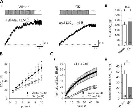
Figure 3.
Initial exocytotic response during repetitive stimulation is impaired in β cells of diabetic rats. (A, i) Typical depolarization train–induced changes in Cm (bottom trace) in a β-cell of a Wistar (left) and GK rat (right). (ii) Comparison of the total ΣΔCm reached after the end of the train between β cells of control (open bars) and diabetic (gray bars) animals. (B) Average ΣΔCm during the first 10 pulses of train stimulation of Wistar (closed circles) and GK (open circles) rat β cells. Note the depressed initial release in diabetic β cells. Straight lines represent linear fits through 10 data points starting with ΔCm evoked by pulse #2. (C) Cell size–normalized cumulative increase in Cm during (i) and at the end (ii) of train stimulation (same data as in A). **, P < 0.01; *, P < 0.05, unpaired t test.

