Figure 3.
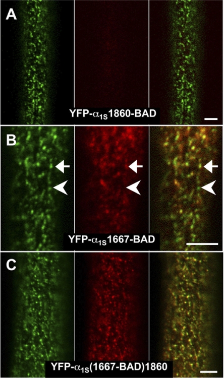
Differential accessibility of α1S C-terminal sites to streptavidin in nonfixed myotubes. (A) Myotubes expressing YFP-α1S1860-BAD did not display binding of streptavidin that colocalized with loci of fluorescent DHPRs. (B) Myotubes expressing YFP-α1S1667-BAD displayed incomplete colocalization between loci of DHPRs and streptavidin binding. The arrow indicates a cluster of DHPRs with substantial binding of streptavidin, and the arrowhead a cluster of DHPRS showing very little binding of streptavidin. (C) Nearly complete colocalization was observed for YFP-α1S(1667-BAD)1860. Bars, 5 μm.

