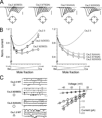
Figure 3.
Mutations at the DCS locus suppress anomalous mole fraction of HVA. The four amino acids of the CaV2.3 DCS locus were mutated by adding or removing negatively charged residues. (A) Typical current traces recorded during voltage ramps under different ionic conditions (see Fig. 1) applied to oocytes expressing CaV2.3 channels with their DCS loci mutated in DSED (one negative charge added, CaV2.3(DSED)), DEEE (two negative charges added, CaV2.3(DEEE)), TSQN, AAAA, or GGGG (two negative charges removed, CaV2.3(TSQN), CaV2.3(AAAA) or CaV2.3 (GGGG)). (B) Current–mole fraction curves obtained for the above mutations. Right, effect of adding negative charges (CaV2.3(DSED), ○; CaV2.3 (DEEE), ▿). Left, effect of removing negative charges (CaV2.3(TSQN), □; CaV2.3 (AAAA), hexagone; CaV2.3 (GGGG), ▿). Note that the removal of the negative charges suppressed AMFE in all three cases. The dotted line represents the curve obtained with the WT CaV2.3 in Fig. 1. (C) Single-channel conductances of CaV2.3, CaV2.3(DSED), CaV2.3(AAAA), and CaV1.2 VGCC. Left, current traces in 100 mM BaCl2 (pipette potential = +10 mV, except CaV2.3(AAAA) −20 mV). Right, current–voltage curves with superimposed linear regressions (13 ± 1 pS, 21 ± 1 pS, 4 ± 1 pS, and 22 ± 1 pS for CaV2.3, CaV2.3(DSED), CaV2.3(AAAA), and CaV1.2, respectively). Bar, 0.5 pA, 25 ms.

