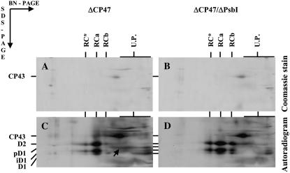
Figure 3.
Analysis of thylakoid membrane proteins in the psbB deletion mutant ΔCP47 and the double deletion mutant ΔCP47/ΔPsbI. Cells of the Synechocystis 6803 strains were radiolabeled at 500 μmol photons m−2 s−1 and 29°C with [35S]Met/Cys for 30 min and their thylakoid proteins were separated by 2D BN/SDS-PAGE. Designations of proteins are as described in the legend to Figure 1; the arrow indicates the complex of PsbI and pD1. Six micrograms of Chl was loaded for each sample.

