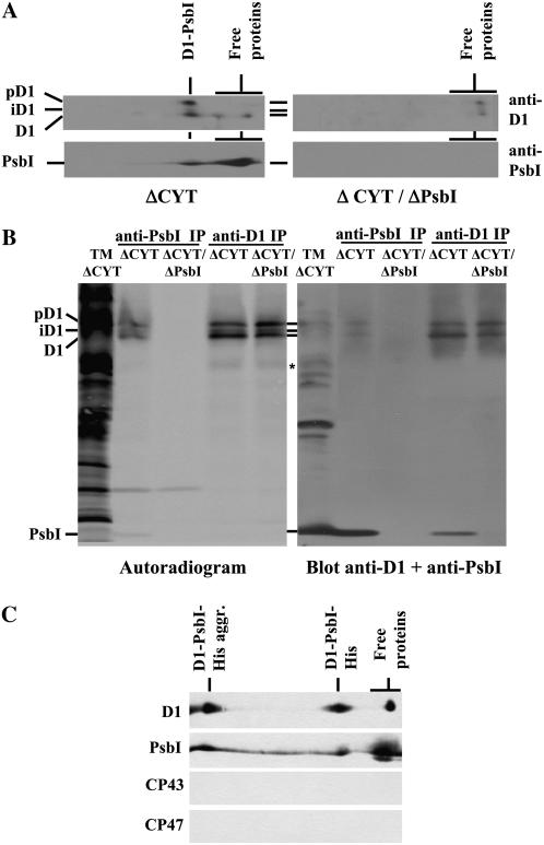
Figure 4.
Identification of a D1-PsbI precomplex. A, Thylakoid membrane proteins (1 μg of Chl) of the ΔCYT and ΔCYT/ΔPsbI strains were separated by 2D BN/SDS-PAGE, transferred onto PVDF membrane, and detected by antibodies against D1 and PsbI. B, Pulse-labeled thylakoid proteins (5 μg of Chl) from the ΔCYT and ΔCYT/ΔPsbI strains were immunoprecipitated using antibodies specific for D1 (anti-D1 IP) or PsbI (anti-PsbI IP) and the immunoprecipitates together with thylakoids from ΔCYT (TM) were analyzed by SDS-PAGE, blotted onto PVDF membrane, autoradiographed (left, autoradiogram), and then probed with antibodies against both PsbI and D1 proteins (right, blot anti-D1 + anti-PsbI). *, A putative 23-kD D1 synthesis intermediate. C, Immunoblots of D1, PsbI, CP43, and CP47 after 2D BN/SDS-PAGE of the protein fraction bound to nickel-affinity column loaded with solubilized thylakoids of the PsbI-His/ΔPsbI/ΔCYT strain. D1-PsbI-His aggr., Aggregates of D1 and PsbI-His.

