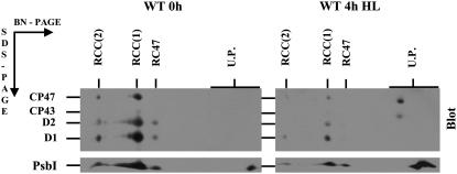
Figure 5.
Immunoblots of thylakoid membrane proteins from the Synechocystis 6803 wild-type strain before and after 4-h exposure to high light. Cells of wild type were exposed to high irradiance (1,000 μmol photons m−2 s−1) for 4 h in the presence of the protein synthesis inhibitor lincomycin (100 μg mL−1). Thylakoid membrane proteins (1 μg of Chl) were separated by 2D BN/SDS-PAGE, transferred onto PVDF membrane, and probed with antibodies against the D1, D2, CP43, CP47, and PsbI proteins. Designations of proteins are as described in the legend to Figure 1. To allow direct comparison of protein bands, thylakoids from control and photoinhibited cells were analyzed on a single gel and blot.

