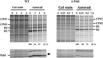
Figure 6.
Degradation of the PSII proteins in the wild-type and ΔPsbI strains under high irradiance monitored by radioactive pulse-chase labeling. Cells of both strains were subjected to 250 μmol photons m−2 s−1 of white light for 20 min in the presence of [35S]Met/Cys. Then the cells were washed, supplemented with unlabeled Met/Cys, and subjected to 500 μmol photons m−2 s−1 of white light for 6 h. Thylakoids were isolated, analyzed by SDS-PAGE, the gel was stained (Gel stain), and the radioactive labeling of the proteins was visualized using a PhosphorImager (Autorad). Quantification of radioactivity in the D1 band was performed by ImageQuant software with samples of each strain equally loaded on Chl basis (2 μg of Chl; see Gel stain) in a single gel. The radioactivity incorporated into the D1 band of each strain during pulse was taken as 100%; numbers show means of three measurements; sd did not exceed 7%. The low-Mr region of the gel is shown in the bottom sections and the stable band of PsbI is designated by an arrow.

