Figure 4.
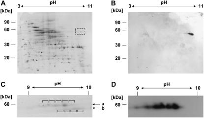
The two DEAD-box proteins are detectable in mitochondria from cell suspension cultures. Total mitochondrial protein is analyzed by IEF (first dimension) and Tricine-SDS-PAGE (second dimension). A, Coomassie staining revealed spots in the area where PMH2 is expected from previous studies (pH 10/60 kD; dashed box). B, Immunodetection analysis with the PMH1/PMH2 antiserum detects specific spots in this area. C and D, Enhanced resolution of the proteins was achieved on pH 7 to 11 gradients. A section of the stained gel (C) shows spots in expected size and pH range (indicated by arrows a and b). Relevant spots (indicated by brackets) were excised from the gel and analyzed by MS. In all spots of the top row, PMH1 and/or PMH2 is the predominant protein. D, Immunodetection analysis indicates the presence of the PMH proteins in the top row of the proteins seen between pH 9 to 10.

