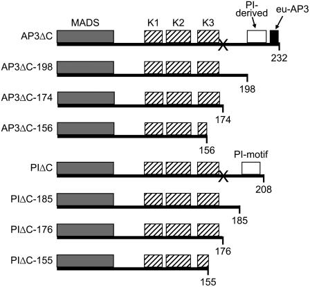
Figure 1.
Summary of constructs. Schematic representation of the constructs used in this study. The gray box indicates the DNA-binding M domain. Hatched boxes indicate the three putative amphipathic α-helices in the K domain (K1, K2, and K3). Positions of the PI-derived (white box) and euAP3 motif (black box) of AP3 and the PI motif (white box) of PI are indicated. Numbers at the bottom represent amino acid positions. “X” represents the position of the engineered stop codon.

