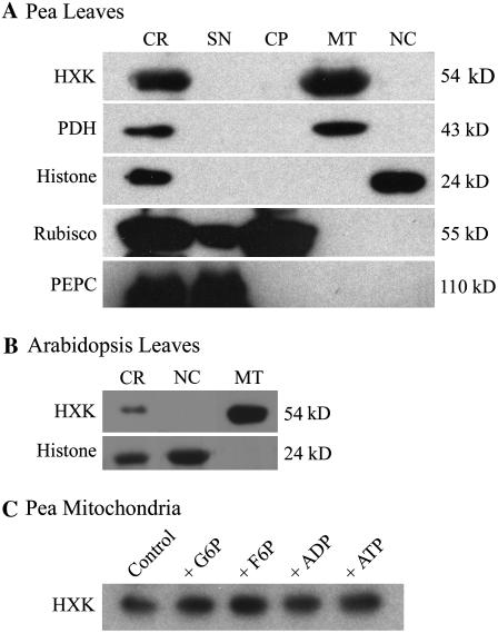
Figure 3.
HXK localization in isolated leaf organelles. A, Western blot of Percoll-purified pea leaf organelles, showing corresponding marker antigens and molecular weights after SDS-PAGE. CR, Crude homogenate; SN, postmitochondrial supernatant; MT, mitochondria; NC, nucleus. B, Western blot of isolated mitochondria and nuclei from Arabidopsis Ler leaves. Leaf organelle fractions also were monitored by fluorescence staining or Chl autofluorescence. These confirmed the purity of pea leaf organelles and also indicated that the Arabidopsis mitochondrial fraction contained low amounts of chloroplasts, whereas the Arabidopsis nuclear fraction was free of mitochondria and chloroplasts (images not shown). C, Western blot for HXK from a washed pea mitochondrial fraction, following overnight incubation at 4°C with 10 mm solutions of indicated metabolites.

