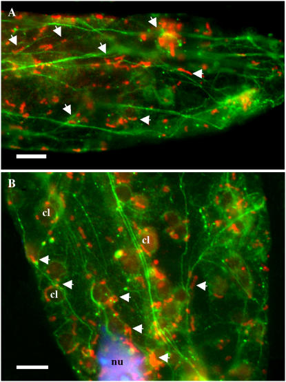
Figure 5.
Association of HXK1-localized mitochondria with F-actin. Following cryofixation and freeze substitution, leaf cells of wild-type (Ler) plants were double labeled with anti-HXK1 polyclonal antibody and antiactin monoclonal antibody. Texas Red- and FITC-conjugated secondary antibodies were used to detect HXK1 and actin, respectively. A and B, Fluorescence images of single optical sections of two representative cells are shown. DNA staining with DAPI is shown in blue (B). Arrowheads show the association of some of the HXK1-labeled mitochondria (red) with actin filaments (green) throughout both fields and with the actin basket surrounding the nucleus (nu) and chloroplasts (cl). Chloroplasts in B show slight autofluorescence. Scale bars = 10 μm.

