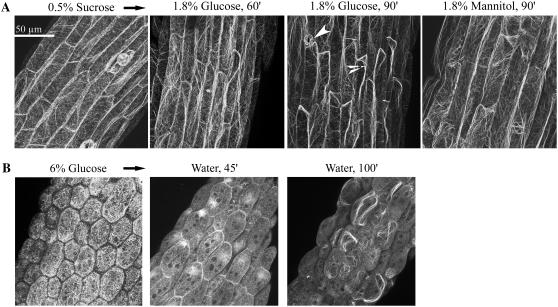
Figure 7.
Glc disrupts F-actin organization as visualized in hypocotyls of GFP-hTalin seedlings. A, Influence of short-term Glc treatment on F-actin organization after seedling transfer to solutions of 1.8% (0.1 m) Glc or mannitol. Prior to treatment, seedlings were grown on agar plates with 0.5% Suc (5 d). Note the loss of many fine filaments after 60 and 90 min of Glc treatment. Arrowheads point to an open stomate and an actin cable. Scale bar = 50 μm. Gain and amplifier offset on the confocal microscope were kept constant throughout the acquisition time courses. Mannitol treatment had no apparent influence on F-actin organization throughout the 90-min treatment time course (earlier images not shown). B, Visualization of actin organization in seedlings developmentally repressed by Glc and the recovery of F-actin after seedling transfer to water. Seedlings were grown on 6% Glc (7 d), then transferred to water for indicated times. These seedlings had arrested growth, with pale white cotyledons. Images are of different seedling hypocotyls. Note the initial absence of fine mesh filaments before transfer and the beginning reorganization of F-actin after transfer to water. Seedlings transferred from 6% mannitol to water showed no apparent changes in their filaments over several hours (observations).

