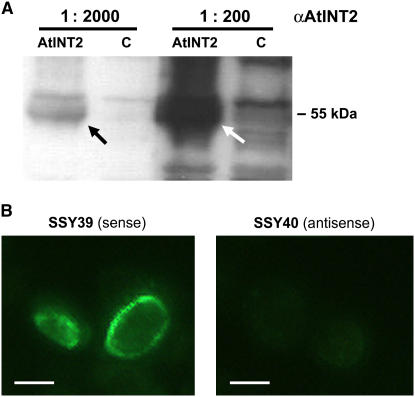
Figure 6.
Immunohistochemical detection of recombinant AtINT2 protein in western blots of yeast total membranes and in thin sections of AtINT2-expressing yeast cells. A, Unpurified αAtINT2 (diluted 1:200 or 1:2,000) that had been raised against 26 amino acids from the AtINT2 C terminus labeled a 55-kD band in detergent extracts from yeast total membranes after gel electrophoresis and blotting to nitrocellulose filters (10 μg lane−1; AtINT2 = AtINT2-expressing cells; C = control cells). B, Incubation of thin sections with affinity-purified αAtINT2 and fluorescence-tagged second antibody yielded fluorescence only in AtINT2-expressing cells (SSY39) but not in control cells (SSY40). Bars are 2 μm. [See online article for color version of this figure.]

