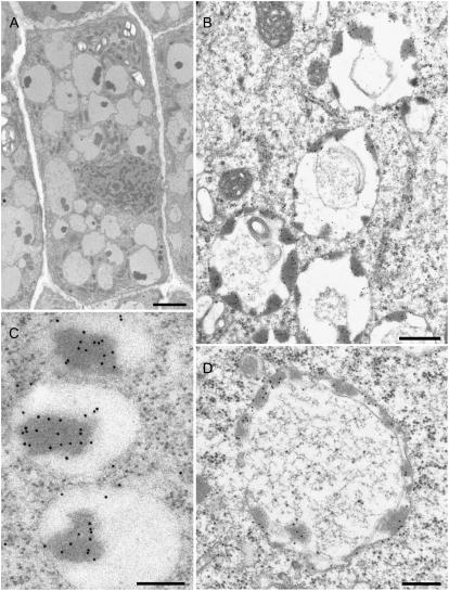
Figure 6.
Immunogold detection of barley lectin in vacuoles of 3-d-old barley roots. A, Cell in the calyptra revealing electron opaque aggregates within the vacuoles. B to D, Electron opaque aggregates stain positively with anti-wheat germ agglutinin gold (10 nm) conjugates. C and D, Stages in vacuole formation in cells immediately bordering on the meristem. Barley lectin-positive aggregates are positioned more on the outer than the inner tonoplast membrane. Bars = 1.5 μm (A), 400 nm (B), 150 nm (C), 300 nm (D).

