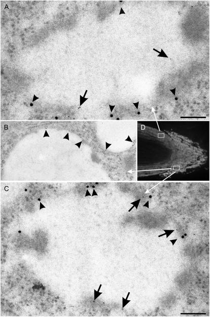
Figure 7.
Immunogold detection of TIPs in cells from 3-d-old barley roots. A and C, Double immunogold labeling with α-TIP (15-nm gold particles, arrowheads; α-TIP peptide antiserum [Jauh et al., 1998]) and γ-TIP (5-nm gold, arrows; γ-TIP peptide antiserum [Jauh et al., 1998]). B, Single immunogold labeling of a PSV (note storage protein aggregates on the tonoplast) with γ-TIP peptide antiserum (10-nm gold [Jauh et al., 1998]). D, Longitudinal section of root for cell positioning. Boxes indicate areas in the cortex and calyptra from which the vacuoles in A and C are depicted. Bars = 100 nm (A and C); 200 nm (B).

