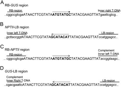
Figure 6.
Sequence analysis of the RB-GUS region (A), NPTII-LB region (B), RB-NPTII region (C), and the GUS-LB region (D). The loxP sequence is indicated in capital letters, and the 8-bp spacer region of the loxP sequence in bold and its orientation by an arrow. The presence of the inverted T-DNA orientation was also clearly demonstrated: the RB-NPTII fragment was composed of the original RB-loxP region and the complement sequence of the inner LB T-DNA region, while the LB-GUS fragment harbored the complement inner RB region and the original loxP-LB region.

