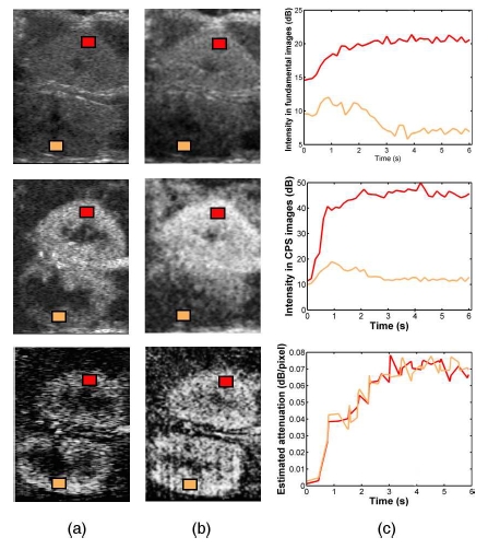
Fig. 3.

Images acquired in fundamental mode (top) and in CPS mode (middle) 2 seconds (a) and 4 seconds (b) after the contrast agent enters the acquisition plane. (bottom) Associated parametric images of microbubble attenuation. (c) Time-intensity curves in both cortices extracted from the fundamental acquisition (top), the CPS acquisition (middle) and the attenuation sequence (bottom).
