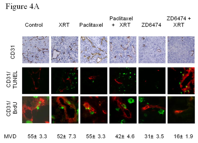
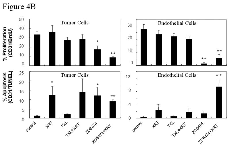
Figure 4.


Microvessel density (MVD) and tumor and endothelial cell proliferation and apoptosis. A, Representative sections obtained from primary lung tumors stained with CD31 (brown staining, upper panels) or double stained with CD31 (endothelial cells, red fluorescence) and TUNEL (apoptosis, green fluorescence, middle panels) or BrdU (proliferation, green fluorescence, lower panels). Proliferating or apoptotic endothelial cells fluoresce yellow and proliferating or apoptotic tumor cells fluoresce green. MVD was quantified in 10 random 0.159-mm2 fields at ×100 magnification. B, Endothelial and tumor cell proliferation and apoptosis. BrdU- or TUNEL-positive endothelial cells were counted in 10 random 0.039-mm2 fields at ×200 magnification. Data was obtained from tumor tissues from 4 or more mice in each treatment group and are plotted as means ± standard error for two independent experiments. *p<0.05, **p<0.01 versus control.
