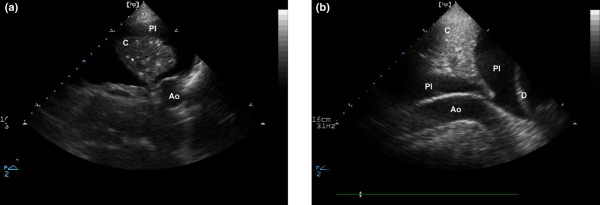
Figure 3.

Ultrasound aspect of a lung consolidation and a pleural effusion. (a) Transversal view of consolidated left lower lobe; lung consolidation is seen as a tissular structure (C). In this consolidation, hyperechoic punctiform images (indicated by asterisk) can be seen; these correspond to air bronchograms (air-filled bronchi). Pleural effusion is anechoic (Pl). (b) Cephalocaudal view of consolidated left lower lobe: lung consolidation with air bronchograms. Ao, descending aorta; D, diaphragm; Pl, pleural effusion.
