
Figure 3.
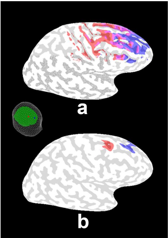
The half-sensitivity areas for the potential (a) and Laplacian (b) due to two electrodes spaced by about 5 cm (shown in the figure inset at left) are plotted using the 3-ellipsoid head model. Half-sensitivity areas are defined as the surface areas of the cortex containing sources that could contribute at least half the “maximum signal” (i.e., contributed by the strongest source location for each electrode or MEG sensor position). The regions of cortex associated with the half-sensitivity area of each electrode are indicated by the two colors. The areas associated with the scalp potential measure are shown to overlap substantially, whereas the scalp Laplacians at these two electrodes are shown to be sensitive mainly to distinct local gyral crowns reflecting the Laplacian's much improved spatial resolution.