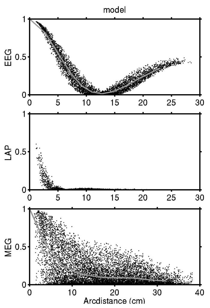
Figure 4.
Simulated coherence is shown as a function of tangential electrode (or MEG coil) separation for EEG, Laplacian, and MEG for uncorrelated cortical sources, that is, spatial white noise at arbitrary temporal frequency. Sensor separation is defined on the simulated scalp surface (ellipsoidal). These coherence fall-offs with sensor separation were calculated using our 3-ellipsoid head model. Simulated cortical dipoles P(r,t) are distributed over the entire folded cortical surface with locations in gyri and cortical folds. P(r,t) is assumed everywhere normal to the local cortical surface, as obtained from one subject's MRI. The grey lines plot the average coherence at each sensor separation.

