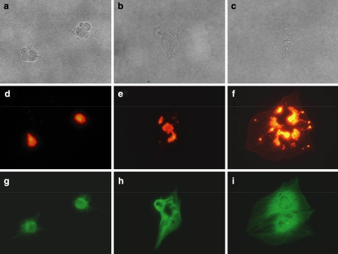
Fig. 3.
Removal of Cuc E on morphology, filament actin, and tubulin of LX-2 cells. Cells were incubated with Cuc E (20 nM) for 24 h and medium was replaced with drug-free medium. Bright field images for (a) 0, (b) 6, and (c) 24 h after medium change. Alexa-phalloidin staining for (d) 0, (e) 6, and (f) 24 h. Tubulin staining for (g) 0, (h) 6, and (i) 24 h

