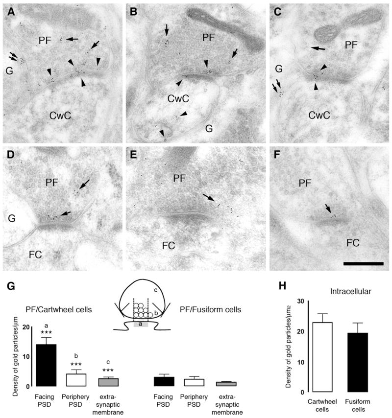
Figure 6. CB1 Receptors Are Differentially Expressed at Parallel Fiber Synapses in DCN.

Electronmicrographs show postembedding immunogold labeling using the CT antibody for CB1 receptor at the parallel fiber (PF)/cartwheel cell (CwC) (A–C) and parallel fiber/fusiform cell (FC) (D–F) synapses. Gold particles for CB1 were observed at the presynaptic boutons (arrows) of parallel fibers synapsing on apical dendrites of FC. Three patterns of immunogold labeling were observed: (1) gold particles associated with the presynaptic membrane, (2) gold particles associated with plasma membrane not facing synapses or extrasynaptic plasma membrane, and (3) gold particles associated with synaptic vesicles intracellularly. In the dendritic spines of cartwheel cells but not fusiform cells gold particles were observed associated with intracellular compartments or plasma membrane (arrowheads). Glial cells (G) also presented gold particles for CB1 (double arrows in [A] and [C]). Scale bar, 0.25 μm. (G) Density of gold particles/mm length ± SEM for CB1 of the parallel fiber synaptic ending plasma membrane. The plasma membrane of the parallel fiber synaptic terminal was divided into three regions, illustrated in the cartoon: parallel fiber membrane facing the PSD (a); parallel fiber membrane facing the periphery of the PSD (b); and parallel fiber membrane not facing the synapse (c) on cartwheel and fusiform cells (***p < 0.001). (H) Density of gold particles/μm2 area ± SEM of the intracellular pool of CB1 in the parallel synaptic terminal onto cartwheel and fusiform cells.
