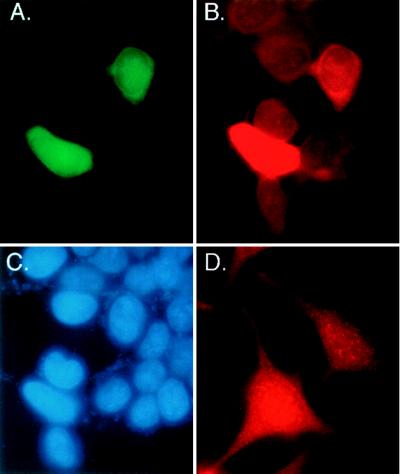
Figure 2.
Localization of mE2 protein in the cytoplasm and nucleus of mammalian cells. Histochemistry showing endogenous mE2 protein and transiently expressed GFP-mE2 fusion protein localizes in both the cytoplasm and nucleus of human cells. (A) Detection of green fluorescence in both the cytoplasm and nucleus of 293T cells expressing GFP-mE2 fusion protein. (B) mE2-like immunoreactivity was detected in red fluorescence. Two cells expressing GFP-mE2 fusion protein and other cells having endogenous-E2 show the same distribution pattern, both in the cytoplasm and nucleus. (C) The same cells used in A and B were stained with 4′,6-diamidino-2-phenylindole to visualize the nucleus. (D) mE2-like immunoreactivity was detected in red fluorescence in the cytoplasm and nucleus of HeLa cells.

