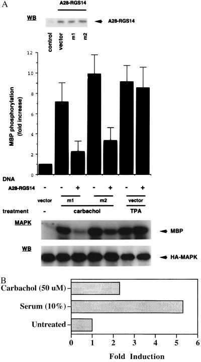
Figure 3.
(A) A28-RGS14 coexpression blocks agonist-induced activation of ERK2 in M1- or M2-transfected COS-7 cells. Plasmids encoding M1 or M2 muscarinic receptors were cotransfected with a pcDNA3-A28-RGS14 cDNA vector or empty expression vector, as indicated, and a plasmid encoding HA-tagged MAPK (ERK2). Cells were exposed to Carbachol (100 μM) or phorbol 12-myristate 13-acetate (100 ng/ml) for 5 min and lysed, and ERK activity was assayed on anti-HA immunoprecipitates. After autoradiography, radioactivity incorporated into myelin basic protein (MBP) was quantitated with a Molecular Dynamics PhosphorImager. Data represent the mean ± SEM of six to eight experiments, expressed as fold increase with respect to vector-transfected cells (control). Fifty micrograms of total lysate proteins was subjected to Western blot analysis using anti-HA or anti-A28-RGS14 mouse monoclonal antibodies (Ab-1C5; Y.C., L.B., and N.K., unpublished results). (B) Induction of A28-RGS14 expression in response to mitogenic signals—a potential role as negative feedback regulator. NIH 3T3 cells expressing the muscarinic M1 receptor (10) were grown to confluence, transferred to serum-free medium (DMEM containing 0.1% BSA) for 16 h, and then stimulated with or without Carbachol (50 μM) or fetal bovine serum (10%) for 6 h. RNA prepared from the samples was examined by Northern blot analysis using 32P-labeled probes for mouse A28-RGS14 and GAPDH (as in Fig. 1A). Autoradiograms were analyzed by laser densitometric scanning (Applied Biosystems) and the signal was normalized to GAPDH control.

