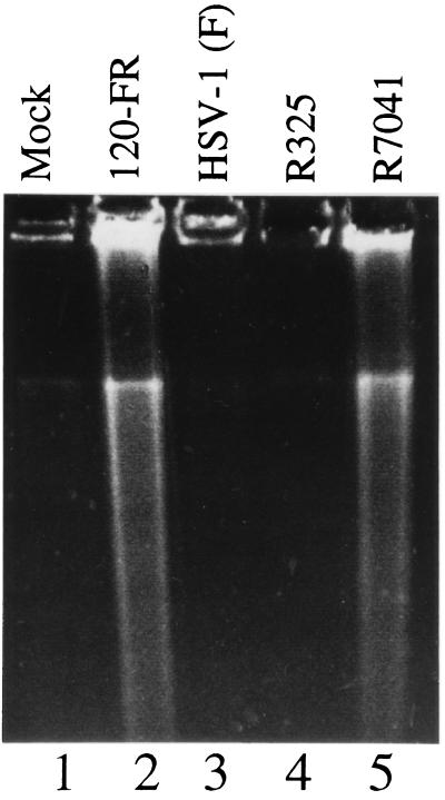
Figure 3.
Photograph of an agarose gel containing electrophoretically separated DNA fragments and stained with ethidium bromide. Vero cells were mock-infected (lane 1), infected with HSV-1 120FR mutant (lane 2), wild-type HSV-1(F) (lane 3), HSV-1 R325 mutant (lane 4), or HSV-1 R7041 mutant (lane 5). Cells were harvested and processed as described in the legend to Fig. 2.

